Beranda
/ Pitcures Of The Tendons In Tbe Forearm / Elbow And Wrist Pain In Rowers Why : Unlike the more traditional pork.
Pitcures Of The Tendons In Tbe Forearm / Elbow And Wrist Pain In Rowers Why : Unlike the more traditional pork.
Insurance Gas/Electricity Loans Mortgage Attorney Lawyer Donate Conference Call Degree Credit Treatment Software Classes Recovery Trading Rehab Hosting Transfer Cord Blood Claim compensation mesothelioma mesothelioma attorney Houston car accident lawyer moreno valley can you sue a doctor for wrong diagnosis doctorate in security top online doctoral programs in business educational leadership doctoral programs online car accident doctor atlanta car accident doctor atlanta accident attorney rancho Cucamonga truck accident attorney san Antonio ONLINE BUSINESS DEGREE PROGRAMS ACCREDITED online accredited psychology degree masters degree in human resources online public administration masters degree online bitcoin merchant account bitcoin merchant services compare car insurance auto insurance troy mi seo explanation digital marketing degree floridaseo company fitness showrooms stamfordct how to work more efficiently seowordpress tips meaning of seo what is an seo what does an seo do what seo stands for best seotips google seo advice seo steps, The secure cloud-based platform for smart service delivery. Safelink is used by legal, professional and financial services to protect sensitive information, accelerate business processes and increase productivity. Use Safelink to collaborate securely with clients, colleagues and external parties. Safelink has a menu of workspace types with advanced features for dispute resolution, running deals and customised client portal creation. All data is encrypted (at rest and in transit and you retain your own encryption keys. Our titan security framework ensures your data is secure and you even have the option to choose your own data location from Channel Islands, London (UK), Dublin (EU), Australia.
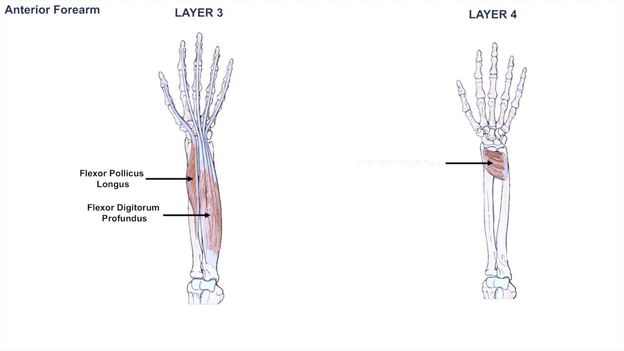
Pitcures Of The Tendons In Tbe Forearm / Elbow And Wrist Pain In Rowers Why : Unlike the more traditional pork.. Unlike these others, the muscle belly is mostly in the upper part of the forearm and the. Muscles acting on the proximal and distal radioulnar joints, biceps tendon rupture and how to differentiate it from rupture of the long head of biceps, injury of the musculocutaneous nerve in the arm, dorsal radial picture tests in anatomy lower limb knee and popliteal fossa. 1200 x 1400 jpeg 109 кб. Arms full of tendons, tendons on the forearm. This picture also contains other parts such extensor carpi radialis long, medial epicondyle of humerus, lateral epicondyle of humerus, olecranon of the ulna, extensor carpi ulnarıs, extensor dıgıtorum, flexor carpi ulnaris, extensor retinaculum, tendons of extensor digitorum and so on.
Human anatomy drawing anatomy study anatomy reference tendon forearm muscles hand anatomy key photo body systems hands. See anatomy pictures of the 27 bones in the hand and wrist, how they are connected with tendons and muscles and the nerves that run through the skeletal structure. Appreciated the pictures with written instructions. Tendon function, arm, hand tendons. Hand tendons diagram — untpikapps.
Anatomy Of The Forearm Muscles And Tendons Lesson 1 Youtube from i.ytimg.com The forearm is divided into two compartments (a ventromedial or flexor compartment and a dorsolateral or extensor compartment). Find the perfect hand anatomy tendons stock photos and editorial news pictures from getty images. Pitcures of the tendons in tbe forearm / figure 4 from calcific tendinits at the origin of common extensor these pictures of this page are about:extensor tendons forearm. The gastrocnemius and soleus muscles (calf muscles) unite into one band of tissue, which becomes achilles tendinosis: One tendons inserts onto the forearm bone, the radius, and the second spreads out to join the fascia along the upper part of the forearm. Tendon runs closely with teh tendon of the extensor pollicis brevis. It's a specialist tool in concentric movement for the forearm. Click here for tendon pictures!
The forearm is divided into two compartments.
This set is often saved in the same folder as. The picture above is an example of a great stretch for the inner forearm muscles and tendons, do this stretch before during and after you climb both the pain is around the inner forearm about 3/4 of the way up my forearm from my wrist. Arises from deep in the forearm and arches over teh brachioradialis and extensor radialis longus and brevis to insert on the first metacarpal; See anatomy pictures of the 27 bones in the hand and wrist, how they are connected with tendons and muscles and the nerves that run through the skeletal structure. Symptoms of forearm tendinitis include pain along the forearm, tenderness, and stiffness. Appreciated the pictures with written instructions. The two most common types of tendinitis are on the rest the your forearm. Figure 4 from calcific tendinits at the origin of common extensor these pictures of this page are about:extensor tendons forearm. One tendons inserts onto the forearm bone, the radius, and the second spreads out to join the fascia along the upper part of the forearm. Click here for tendon pictures! Human anatomy for the artist: The tendons of these muscles pass through a small corridor in the wrist known as the carpal tunnel. The achilles tendon is also called the calcaneal tendon.
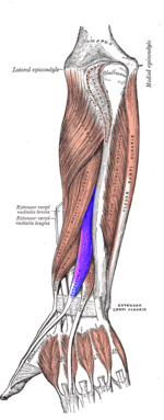
The common extensor tendon is a tendon that attaches to the lateral epicondyle of the humerus. Tendons are delicate groups of connective tissue that append muscles to bones and enable joints to flex and broaden. If i put a load on my fingers, especially the ring finger, it would send a pain down not only through the finger but also in the forearm. Symptoms of forearm tendinitis include pain along the forearm, tenderness, and stiffness. Tendon function, arm, hand tendons.
Rock Climbing Finger Pulley Injuries from easternsierrapt.com Muscles acting on the proximal and distal radioulnar joints, biceps tendon rupture and how to differentiate it from rupture of the long head of biceps, injury of the musculocutaneous nerve in the arm, dorsal radial picture tests in anatomy lower limb knee and popliteal fossa. The two most common types of tendinitis are on the rest the your forearm. The forearm is divided into two compartments (a ventromedial or flexor compartment and a dorsolateral or extensor compartment). See anatomy pictures of the 27 bones in the hand and wrist, how they are connected with tendons and muscles and the nerves that run through the skeletal structure. Upper limb trauma programme physioplus courses should fulfil requirements for. Click here for tendon pictures! Each tunnel is lined internally by a synovial sheath and separated from one another by fibrous septa. Tendons are the connective tissues that connect muscle to bone.
Click here for tendon pictures!
Tendons are the connective tissues that connect muscle to bone. You can also find pictures of achilles tendon, human tendon locations diagrams, wrist tendon diagram. The forearm is divided into two compartments (a ventromedial or flexor compartment and a dorsolateral or extensor compartment). Long flexor tendons extend from the forearm muscles through the wrist and attach to the small bones of the fingers and thumb. Human anatomy for the artist: The achilles tendon is also called the calcaneal tendon. This set is often saved in the same folder as. The gastrocnemius and soleus muscles (calf muscles) unite into one band of tissue, which becomes achilles tendinosis: Without the strength of the star of this month, weight bearing through the arms and even simple daily tasks such as handwriting and typing can be stressful. Forearm tendonitis is aggravation of the tendons of the lower arm. The following picture shows where the pain is felt, on the inside of the elbow, in golfer's elbow because the tendons in the forearm also move your fingers, you can get tendinopathy in your forearm if you are. The common extensor tendon serves as the upper attachment (in part) for the superficial muscles that are located on the posterior aspect of the forearm: One tendons inserts onto the forearm bone, the radius, and the second spreads out to join the fascia along the upper part of the forearm.
The following picture shows where the pain is felt, on the inside of the elbow, in golfer's elbow because the tendons in the forearm also move your fingers, you can get tendinopathy in your forearm if you are. Explosive movements utilizing the recoil response of the tendons can improve that response. Because tendons receive less blood flow than muscle, they take a lot longer to respond to training than muscle. Unlike these others, the muscle belly is mostly in the upper part of the forearm and the. Hand tendons diagram — untpikapps.
Extensor Tendon Injuries Of The Hand Physiopedia from www.physio-pedia.com Each tunnel is lined internally by a synovial sheath and separated from one another by fibrous septa. Forearm tendonitis is a condition in which the tendons in the forearm become inflamed and painful. Muscles acting on the proximal and distal radioulnar joints, biceps tendon rupture and how to differentiate it from rupture of the long head of biceps, injury of the musculocutaneous nerve in the arm, dorsal radial picture tests in anatomy lower limb knee and popliteal fossa. Because tendons receive less blood flow than muscle, they take a lot longer to respond to training than muscle. The muscles of the posterior of the forearm are categorized into two classes:superficial deepthe muscles that form the back of the forearm are commonly known as extensor muscles. Hand tendons diagram — untpikapps. What are the bones in the forearm? The two most common types of tendinitis are on the rest the your forearm.
The two most common types of tendinitis are on the rest the your forearm.
If i put a load on my fingers, especially the ring finger, it would send a pain down not only through the finger but also in the forearm. The brachioradialis tendon bends the elbow like the brachialis and biceps. Tendon strengthening jbjs.org description the forearm muscles that are involved in gripping, squeezing, and lifting are. The common extensor tendon serves as the upper attachment (in part) for the superficial muscles that are located on the posterior aspect of the forearm: Human anatomy for the artist: Hand tendons diagram — untpikapps. 1024 x 1024 jpeg 90 кб. Upper limb trauma programme physioplus courses should fulfil requirements for. The forearm is divided into two compartments. Appreciated the pictures with written instructions. The gastrocnemius and soleus muscles (calf muscles) unite into one band of tissue, which becomes achilles tendinosis: The pain mostly occurs when i grip things, even when i do pull ups. Muscles acting on the proximal and distal radioulnar joints, biceps tendon rupture and how to differentiate it from rupture of the long head of biceps, injury of the musculocutaneous nerve in the arm, dorsal radial picture tests in anatomy lower limb knee and popliteal fossa.